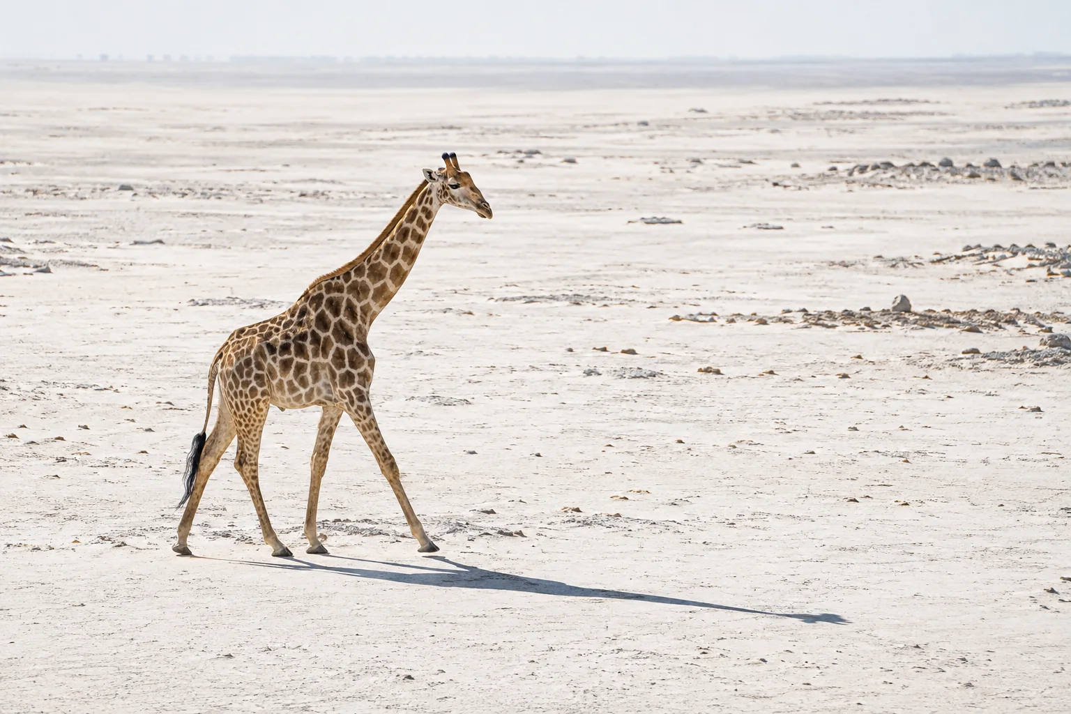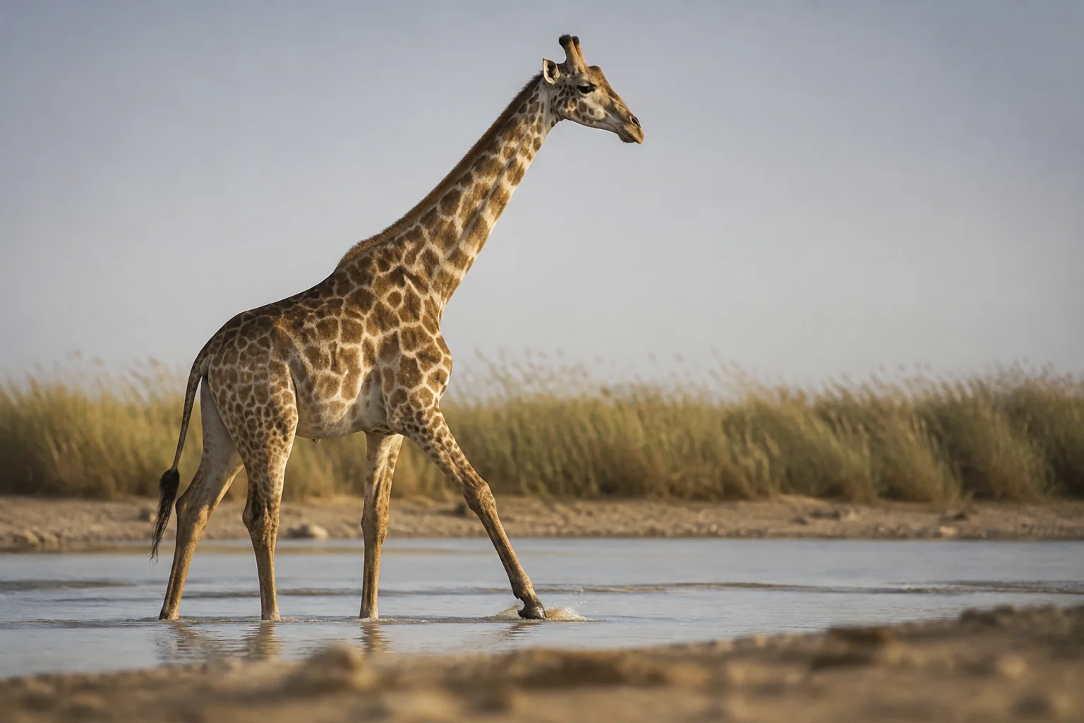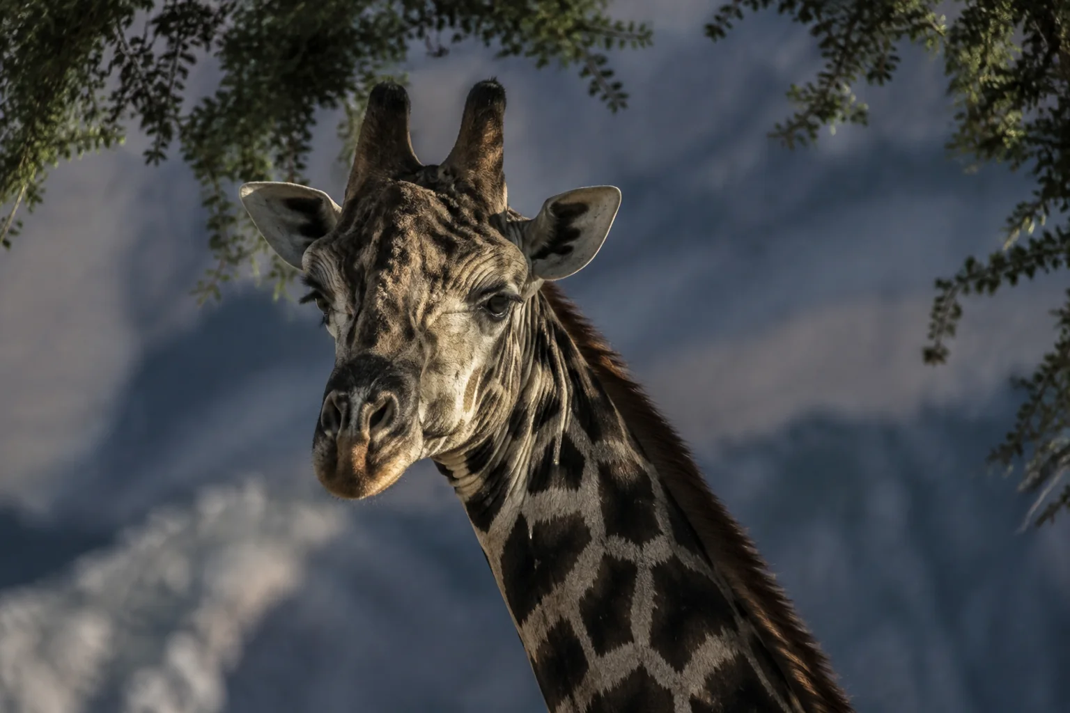
A giraffe walks past, head high, with no obvious wound. Does that mean it is healthy? In one study, X-rays found mild foot-bone changes in three apparently healthy wild giraffes. The lesson is not that every animal conceals an emergency. It is that appearance, movement and a clinical investigation answer different questions—and confusing them can make us miss a problem or invent one.
What does giraffe health include?
Health is more than the absence of a visible injury. For this guide, the useful questions are whether a giraffe can obtain food, maintain its condition and move normally, and whether injury or disease warrants professional investigation. A striking rescue is one part of that picture; repeated observations and less visible diagnostic work matter too.
There are also two different scales. Individual care asks what is happening to a particular animal and what response is appropriate. Population surveillance asks where health problems occur, how often they are detected and whether the pattern changes. A successful individual treatment does not, by itself, demonstrate population recovery.
The sections below explain those distinctions through specific studies and professional guidance. They are not a guide to diagnosis, feeding, handling or treatment. Start with what giraffes eat for their browser diet; the photographic body-condition review explores one monitoring method in depth.
Food, water and body condition
A browser is not just a tall grass-eater
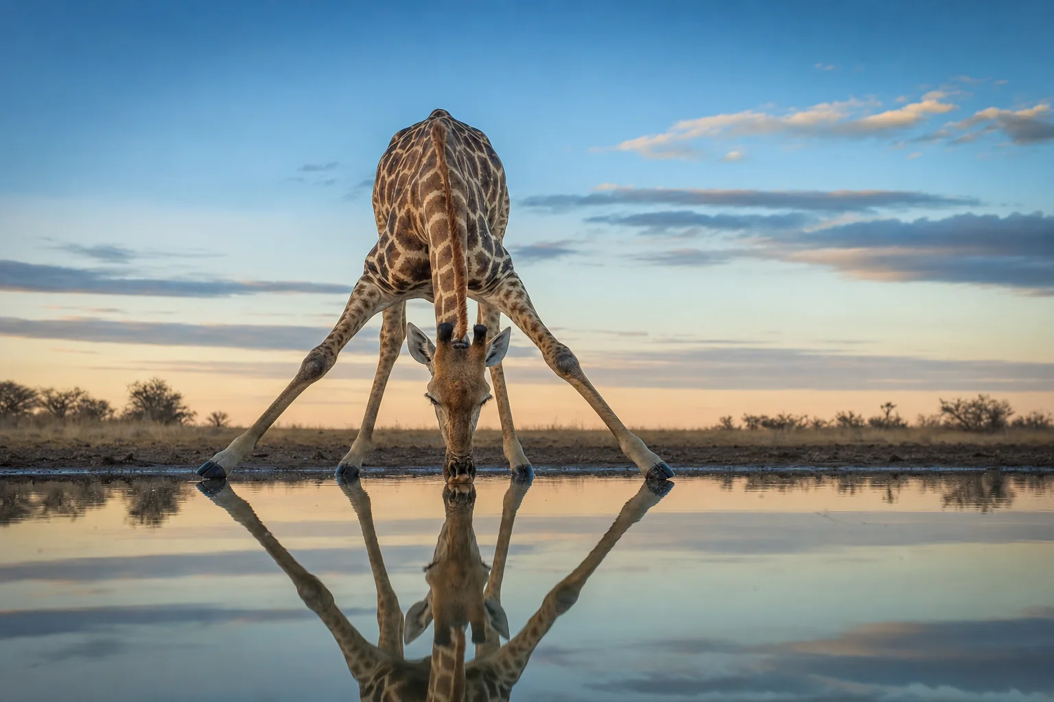
Giraffes browse woody vegetation, particularly leaves, and are ruminants: swallowed food returns to the mouth for further chewing. Moisture in leaves contributes to their water intake. That helps explain why a giraffe's relationship with drinking water differs from ours; it does not mean water is unnecessary or that a fixed drinking interval applies everywhere. These basic adaptations are described in the San Diego Zoo Wildlife Alliance giraffe account.
Food becomes a health question through a chain of evidence: suitable browse is available; the giraffe can reach and consume it; its condition is followed over time. Each observation adds something. If condition changes despite apparently plentiful food, that is a question for professional investigation, not proof that adding more food is the answer. A visitor feeding encounter shows only a small part of an animal's diet.
What can a condition photograph establish?
A 2021 study by Clavadetscher and colleagues developed a seven-level score using visible contours around the backbone and hips. Suitable rear or rear-oblique views could provide a condition signal; vegetation, angle and light often made other images unsuitable.
The score orders appearances. It is not a measurement of kilograms or body fat, and does not explain why a low score occurred. Even its reported consistency chiefly concerned the same observer scoring images again, not independent clinicians agreeing on a diagnosis.
Repeated scoring can nevertheless reveal useful patterns. In that study's northern Tanzanian wild sample, hip scores were higher after the long wet season than after the dry season, especially among adult females. That gives researchers a seasonal condition pattern to investigate, rather than a diagnosis from one picture. Comparable views, dates and animal identities make a “before and after” comparison more informative.
Feet and walking: what can be hidden from view?
The foot example behind our opening comes from Dadone and colleagues' 2021 study. During a planned 2017 conservation translocation in Uganda, researchers obtained foot radiographs from 24 apparently healthy young Nubian giraffes. Three had mild radiographic changes of the distal phalanx—the bone within the hoof—classified in the study as pedal osteitis.
These were selected animals from one population, not a random survey of every wild giraffe. Field conditions also limited the imaging views. The finding cannot tell us the prevalence of foot disease across Africa, and it does not prove that all wild feet are healthy or that zoo feet are unhealthy.
Its useful contribution here is narrower: surface appearance and an X-ray can reveal different things. Watching a walk, inspecting the hoof and imaging the underlying bone provide complementary information. The radiographic finding still needs clinical interpretation; it is not automatically a measure of pain or an explanation of how the animal moves.
Skin marks, wounds and disease
Disease means a disruption of normal health or function; it is not always infectious. An infection occurs when an infectious agent establishes in or on a host, while an injury is physical damage, such as a wire-snare wound. These categories can interact, but they answer different questions about what is happening and why.
Giraffe skin disease, or GSD, describes skin conditions with visible lesions, often crusted or scab-like. Their location varies: limbs, chest and neck have all been described. Bernstein-Kurtycz and colleagues' 2023 paper describes this variation, but does not establish a single cause or transmission pathway for every such lesion. An ordinary photograph cannot resolve that question.
The researchers investigated a different question: walking. They compared adult male Nubian giraffes with different visible skin-disease and wire-snare-wound categories. Some wound-group comparisons showed movement differences. Mostly neck-located GSD was not associated with the measured limb variables in that study.
That is a useful functional finding for those animals, not a declaration that every form of GSD is harmless. It shows why lesion location and the outcome measured belong beside a health claim. The skin disease, snare wounds and gait review explains the separate results and their limits.
How do professionals assemble the evidence?
Health work is not a choice between a camera and a vet. Different evidence helps answer different questions. This comparison is our editorial synthesis of the studies and guidance linked here, not a diagnostic checklist.
| Question | Useful evidence | Important limit |
|---|---|---|
| Has visible condition changed? | Comparable, identified photographs over time | Appearance alone does not establish the cause |
| Does movement differ? | Systematic walking observations or video measurements | Movement does not directly measure pain or survival |
| What might explain a concern? | An appropriate professional examination, imaging or laboratory investigation | Results need clinical and sampling context |
| Are more animals affected? | A defined surveillance population, consistent detection and dates | More reports can reflect more observation, not more disease |
| Did an intervention help? | Follow-up with a stated outcome and an appropriate comparison where feasible | One successful case cannot establish a population-wide effect |
The 2024 WOAH–IUCN wildlife surveillance guidelines start with objectives. Targeted surveillance and reports of sick or dead animals can complement each other. Observation, sampling and investigations have different costs, practical limits and welfare considerations; no single method is always the cheapest, safest or sufficient option.
From a concern to follow-up
Across the examples in this guide, a useful way to follow the work is:
- Record the concern. What changed, when, and in which identified animal or group? Responsible staff assess the urgency.
- Define the question. The qualified team decides what assessment or diagnostic evidence is needed. For one giraffe this may concern an individual problem; surveillance asks whether a wider pattern exists.
- Choose a response. The team weighs the evidence, welfare and risks when deciding on observation, intervention or other risk controls. This sequence is an explanation, not a treatment protocol.
- Follow the outcome. Record what happened afterwards. Individual care follows the animal; surveillance follows a defined population and its observation effort.
Investigations of dead animals can also contribute evidence; they are not non-invasive observation of a living giraffe. Captive findings cannot replace free-ranging surveillance. And ten reported cases instead of five may reflect a bigger search rather than more disease—the detection problem explored in our giraffe population survey guide.
Zoo care and voluntary participation
Known animals in managed care can sometimes learn to participate in examinations. A 2022 report by Booth and colleagues describes training a 22-year-old female reticulated giraffe at Dubai Safari Park for voluntary front-foot radiographs.
The giraffe could walk away. Staff watched for discomfort and adjusted their expectations or ended sessions. The case illustrates how sustained keeper–animal work can make some diagnostic access possible without physical or chemical restraint during the trained procedure.
It is one animal in one institution, not a controlled trial or a universal success rate. The value lies in showing the preparation behind diagnostic care: a familiar animal, repeated work and staff responding to its behaviour. That opportunity differs from encountering a free-ranging giraffe. It is not an invitation for visitors to attempt contact or training.
Why moving giraffes needs a health plan
Moving an animal joins the health questions of an individual, a source population and a destination. The IUCN conservation translocation guidelines therefore treat disease risk in context. The aim is not an impossible promise that a moved animal carries no parasites or pathogens. It is a proportionate assessment of relevant risks and benefits.
Screening and quarantine are not interchangeable with a guarantee. IUCN also notes that quarantine decisions need case-specific assessment, including the stress they may introduce. These are general conservation principles, not giraffe-specific instructions or a checklist a visitor can apply.
For a public account of giraffe translocation science, ask what the move was intended to achieve, who was responsible and what follow-up was reported. “The giraffe arrived” answers a transport question. It does not, by itself, answer how it fared afterwards or whether the conservation objective was met.
Giraffe anatomy is distinctive, as the heart and blood-pressure guide explains. Handling and clinical decisions belong with specialist teams; this page provides no immobilisation or treatment instructions.
How to read a health-conservation claim
A credible story helps the reader distinguish observation, interpretation, action and outcome. These five questions make that distinction practical:
- What was observed? A photograph, a change in movement, a laboratory result and a diagnosis are not interchangeable.
- Who was assessed? One identified giraffe, a selected group and a defined wild population support different conclusions.
- What was done, and by whom? Look for the responsible qualified team and a clear purpose, not a dramatic image standing in for an explanation.
- What happened afterwards? Ask which outcome was followed, for how long and whether animals were lost to follow-up.
- What remains uncertain? A report earns trust by stating what it cannot establish, including whether an individual result generalises.
This does not diminish a successful rescue. It lets an individual welfare benefit stand on its own terms without inflating it into evidence of species recovery. Health work also sits alongside the wider threats to giraffes; it is not a replacement for understanding habitat, human pressures and population trends.
What to remember
- Food, condition, movement, injury and disease provide connected but distinct health questions.
- A photograph or walking video can contribute evidence without supplying a diagnosis.
- Small studies and single-animal care reports are useful when their limits stay attached.
- Population health requires defined observation effort and follow-up, not just a tally of memorable cases.
- Veterinary interventions belong with qualified teams; readers can evaluate claims without attempting care themselves.
Frequently asked questions
Can you tell whether a giraffe is healthy from a photograph?
Not completely. A suitable image may document a visible injury or support a condition score, but cannot reveal every problem or explain its cause. Comparable repeated images and other evidence are more informative than judging an animal from one striking picture.
What health problems do giraffes face?
Documented research includes foot-bone changes, visible skin disease and wire-snare injuries; nutrition and body condition are also subjects of health assessment. This is not an exhaustive disease list or a ranking of causes of death. Their importance depends on the animals, place, period and evidence being studied.
Can giraffes take part in veterinary checks without sedation?
Some can participate in trained procedures in managed care, as the single-animal voluntary X-ray case above demonstrates. That does not mean every examination, animal or setting allows the same approach. The responsible veterinary and care teams decide what is appropriate.
What should I do if I see an injured giraffe?
Keep your distance and alert the park authority, guide or zoo staff responsible for the animal. Do not approach, feed, handle or try to treat it. Our how to help giraffes guide focuses on appropriate public actions and checking conservation claims.
Sources
The linked evidence comprises the Dadone foot-radiograph study (2021), Clavadetscher photographic-condition study (2021), Booth voluntary-care case (2022), Bernstein-Kurtycz gait study (2023), San Diego Zoo Wildlife Alliance's diet account, IUCN translocation guidance (2013), and WOAH–IUCN surveillance guidance (2024). Links appear beside the relevant discussion so readers can distinguish primary studies from general professional guidance.
Reviewed 10 September 2026. This general-reader synthesis is not veterinary advice or an exhaustive clinical review. Explore more source-led explanations in the giraffe science hub.