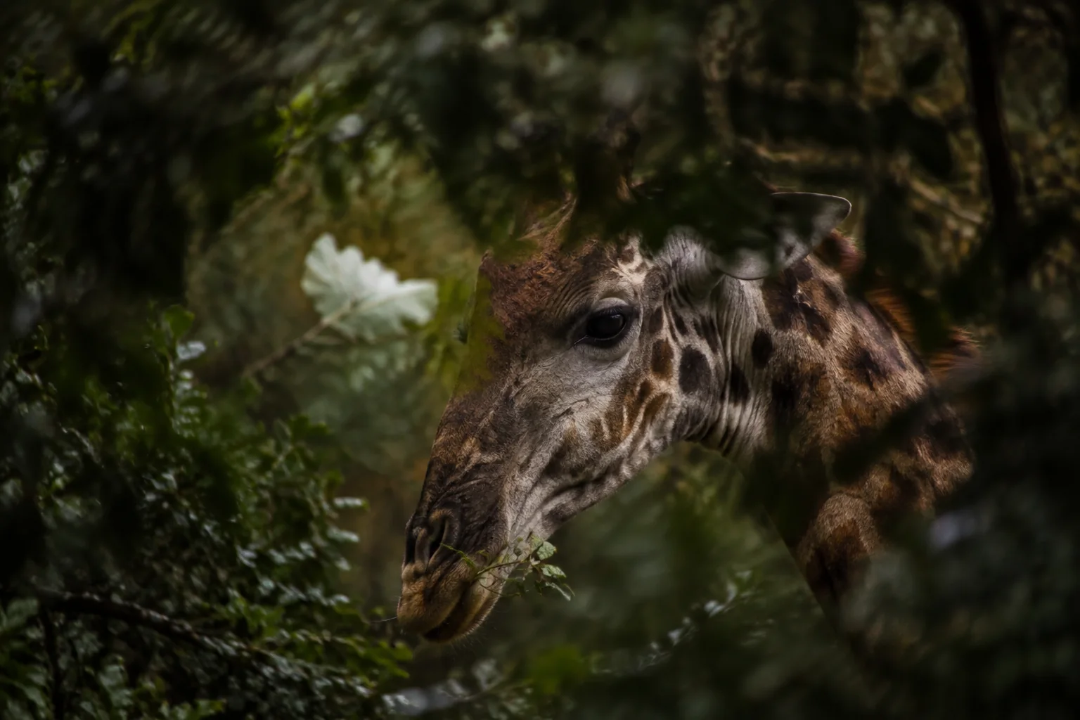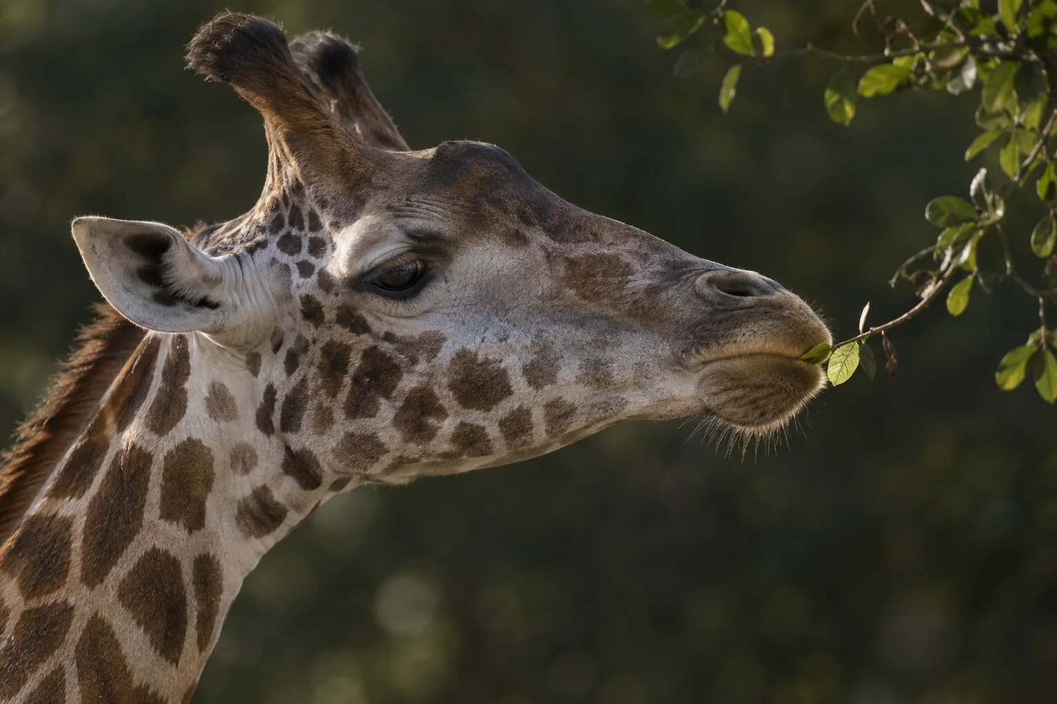
A giraffe’s lips and tongue are visible whenever it takes food. The salivary glands that service its mouth are mostly hidden, as are the ducts carrying their secretions. To understand their arrangement, watching the animal from outside is not enough.
William Pérez, Virginie Michel, Hassen Jerbi and Noelia Vazquez used dissections to describe that anatomy in a 2012 study. They found a broadly familiar ruminant mouth with several distinctive details. One of the most useful comparisons concerned the parotid gland near the rear of the jaw: its shape and the place where its duct delivered saliva differed from the standard cattle anatomy.
Following the structures beneath the skin
The researchers examined the heads of two adult giraffes and one fetus from a zoological collection in Uruguay. They removed the skin, identified facial muscles, then followed salivary glands and their ducts before examining the floor and roof of the mouth. Photographs, radiographs and drawings documented how the parts fitted together.
This was a small descriptive study. The measurements and weights given in the paper came from one of the giraffes, so they should not be treated as averages for the species. The authors also watched two living giraffes feed to connect visible superficial structures with the anatomy they had exposed.
For comparison, the team used established descriptions of cattle anatomy. That was a practical way to identify a shared basic arrangement, but it was not a matched experiment dissecting giraffes and cattle of comparable age and body mass. The study’s strength lay in mapping structures, not in statistically estimating every difference between the species.
A gland shaped around a neighbour
The parotid gland occupied the rear part of the head. In the specimens described, it had two lobes joined below and separated above, creating space for the parotid lymph node. The researchers likened that arrangement to a V around the node.
A lymph node and a salivary gland are different structures. The gland produces secretion delivered into the mouth; the node belongs to the immune and lymphatic system. Identifying their spatial relationship matters because a swelling in the same general region could involve either tissue.
The parotid duct passed forward across the jaw region in a route broadly comparable with cattle. Its destination differed, however. In the giraffe, the authors traced the opening to the region opposite the second upper premolar. The cattle comparison placed the opening farther back, opposite the second upper molar.
That is a difference in the location of delivery, not proof that the giraffe secretes more saliva or has a different saliva composition. Those questions require measurements of secretion and chemistry. The anatomical map tells researchers where the relevant structures are and how they connect.
Several sources of saliva share a narrow space
The mandibular gland was also divided into lobes. One lay between the lower jaws, while the other was partly hidden beneath the parotid gland. Looking only at the exposed surface would therefore miss part of its extent.
Beneath the tongue, the team identified two forms of sublingual gland. One drains through a main duct; the other has multiple small openings. In the giraffe specimens, the former lay farther forward and met its counterpart from the opposite side in the narrow space between the jaws. The latter extended farther back at a deeper level.
Additional salivary tissue occurred in upper, lower and intermediate groups along the cheek. The result was a distributed system rather than a single gland doing all the work. The accompanying drawings are valuable because they show different tissue layers separately: a gland hidden in one view becomes apparent in the next.
These relationships extend the story beyond the giraffe tongue’s visible movements. The mobile organ operates within a mouth whose secretory structures, muscles and passageways must all occupy the available space.
Similar plan, different proportions
The wider mouth examination reinforced the combination of shared structure and specialised shape. The palate was long and narrow, and the tongue’s supporting muscles were strongly developed. The authors identified the familiar ruminant dental arrangement and several muscle groups also described in cattle.
Their conclusion was not that the giraffe possessed an entirely different kind of mouth. Much of its organisation followed the ruminant pattern, while dimensions, mobility and the shapes or positions of particular structures differed. That is a useful anatomical result because it identifies where knowledge from another ruminant may be informative and where direct giraffe observations are needed.
The study did not measure saliva output, chewing forces, gland activity or the protective properties of oral tissues. Nor could three heads establish the range of normal variation across giraffe species, ages and sexes. The authors called for further work on blood supply and innervation, as well as comparisons involving wild animals.
Those questions now have a more explicit anatomical starting point. A future investigation of salivary function can build on mapped glands and ducts rather than assuming their positions from cattle. Pérez and colleagues made the hidden architecture visible, and showed why a familiar-looking arrangement still deserves species-specific description.
Sources
William Pérez, Virginie Michel, Hassen Jerbi and Noelia Vazquez (2012). Anatomy of the Mouth of the Giraffe (Giraffa camelopardalis rothschildi). International Journal of Morphology 30(1), 322–329. DOI; full paper PDF.