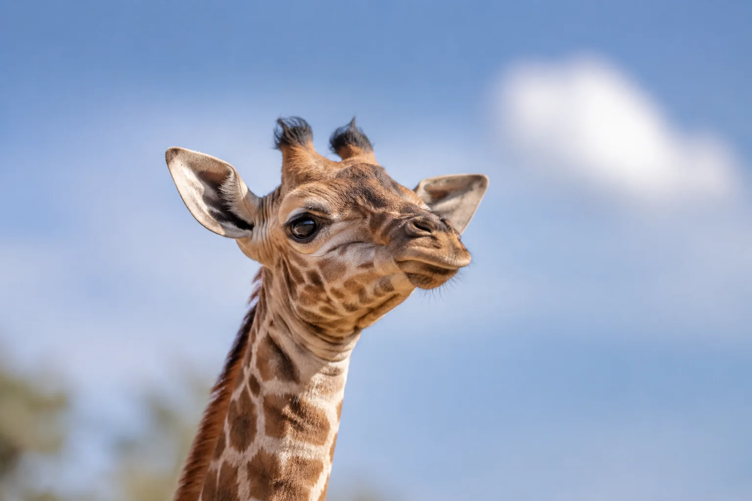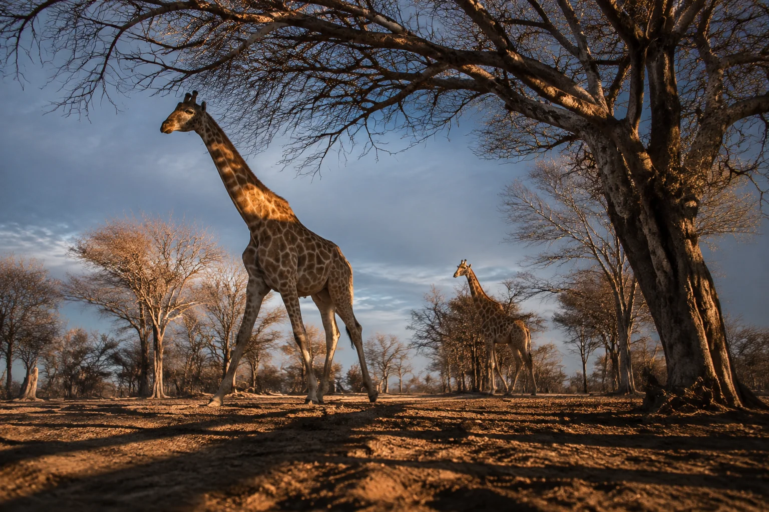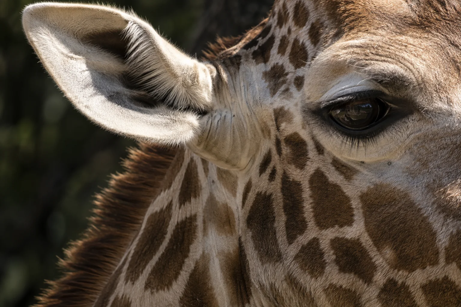
Raising a camera gives it a different view. Turning it changes that view again. A growing giraffe presents both questions: what becomes visible from greater height, and how does the direction of its eyes change?
In a 2013 anatomical study, Graham Mitchell and colleagues examined giraffes culled in Zimbabwe. This was a comparison among differently sized animals, not repeated measurements of the same calves. As faces broadened, the bony eye sockets pointed more towards the front. From those angles, the researchers calculated greater overlap between the two eyes’ estimated views.
What overlapping views contribute
Binocular overlap is the region that both eyes can see. It differs from the entire visual field, which also includes areas visible to only one eye.
Picture two cameras with wide lenses, mounted side by side. Point them apart and each covers more of a different side of the scene. Turn them towards the front and more of their images overlap. The combined panorama and the shared view need not increase together. One can expand while the other contracts.
The shared region makes a particular kind of distance information available. Because the viewpoints are separated, an object appears in slightly different positions relative to its background in the two images. You can see this by holding up a finger and looking through one eye, then the other: the finger appears to shift against the more distant scene.
Comparing the two eyes’ images can support stereoscopic depth perception. But overlap is an opportunity for that comparison, not a measurement of how a particular animal’s nervous system uses it. Distance can also be judged from other information, including perspective and changes as the observer moves.
Nor is depth perception the same as visual acuity. Acuity concerns distinguishing fine detail: whether two nearby points can be seen as separate. Seeing an object’s position in depth and resolving a tiny feature on its surface ask different things of a visual system. A larger shared field should not simply be translated into “sharper eyesight.”
Reading a skull as evidence about sight
The study estimated overlap from the direction of the orbits relative to the skull’s centre line, assuming that this represented the eyes’ optical direction. It did not map the visual fields of living giraffes or test their distance judgments.
That measurement choice explains both the discovery’s value and its boundary. Bones can preserve a structural arrangement; they cannot record every change in gaze. A diagram based on fixed directions is a model of the viewing geometry, rather than a photograph of everything an animal sees at a particular instant.
To investigate an advantage during feeding, a behavioural study would need to connect viewing conditions with performance—for example, whether a giraffe selects or reaches a target more accurately when both eyes can see it. Comparing skull shapes alone cannot answer that question.
The anatomical finding nevertheless adds something concrete to the familiar image of a calf growing taller. The viewing arrangement changes as well as the viewpoint. Understanding giraffe sight therefore involves several connected questions: where the eyes face, how the retina samples an image, and how visual information guides behaviour. Research on giraffe and okapi vision genes addresses another part of that system, the molecules involved in receiving light.
Sources
Mitchell, G., Roberts, D. G., van Sittert, S. J., and Skinner, J. D. (2013). Orbit orientation and eye morphometrics in giraffes (Giraffa camelopardalis). African Zoology, 48(2), 333–339. DOI: 10.1080/15627020.2013.11407600. University of Pretoria record and full paper.