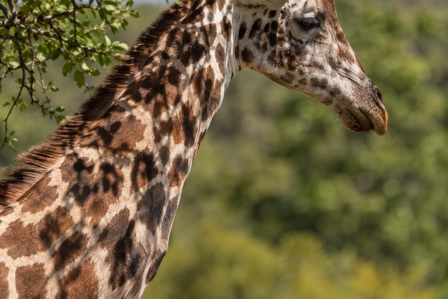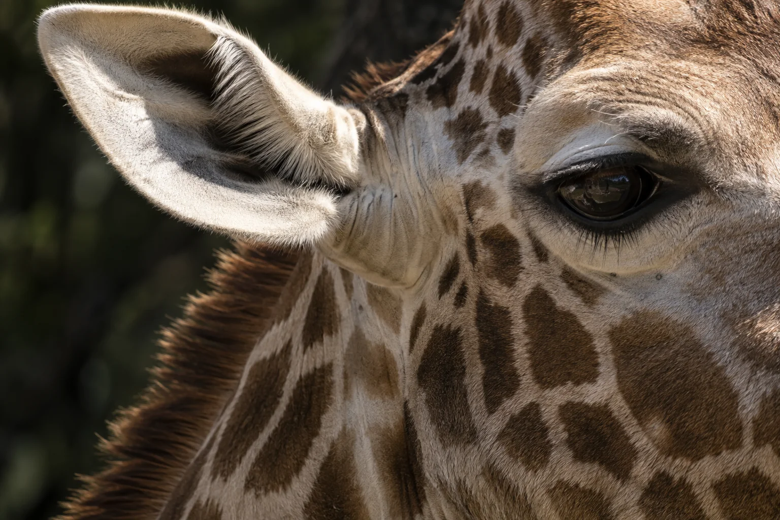
A giraffe and an okapi encounter very different views, but how much of that difference is built into their eyes? Their surroundings alone cannot answer the question. An eye must transmit light, focus it, absorb it in the retina and turn that absorption into a signal. Evolution could modify any of those stages.
Edson Ishengoma, Morris Agaba and Douglas Cavener approached the problem by comparing the DNA instructions for 36 proteins involved in vision. Their 2017 study found that most of these genes were strongly conserved. Against that background, several stood out as candidates for evolutionary change, including a giraffe light-sensitive pigment and an okapi protein involved in the cornea.
The most concrete prediction concerned the giraffe pigment: its sequence suggested a small shift toward absorbing longer wavelengths than the corresponding pigment in the other ruminants examined. That is a specific, testable finding. It is also several steps removed from showing that giraffes recognise a particular colour better or spot predators more effectively.
Looking for changes in a largely conserved system
The researchers began with genes whose involvement in vision was supported by functional evidence. They identified corresponding sequences in giraffe, okapi and other mammals, aligned their protein-coding regions and reconstructed their evolutionary relationships. Comparing relatives helps distinguish a change confined to one lineage from an older feature that several species inherited together.
Some DNA substitutions leave the protein unchanged; others replace one amino acid, a building block of the protein, with another. The balance between those types of substitution can reveal how strongly evolution has constrained a gene. A protein that must retain a precise structure can tolerate very few amino-acid changes.
Across the 36 genes, that constraint was the dominant signal. The study did not uncover wholesale reinvention of the visual system. The authors then used models that allowed particular evolutionary branches or subsets of sites to differ from the general pattern. These tests asked where a small number of changes might deserve closer investigation.
Different statistical results have different meanings here. Evidence of positive selection suggests that amino-acid replacements were favoured in a lineage. Evidence that selection differs between lineages can also reflect stronger preservation of a protein's existing function. Neither statistical label tells the researchers exactly how an eye performs.
A change in the light-catching machinery
The gene OPN1LW encodes an opsin used in a visual pigment sensitive to longer wavelengths. A visual pigment absorbs across a range of wavelengths, with a peak at which absorption is greatest. Changing the surrounding protein can shift that peak without creating an entirely new kind of receptor.
In the giraffe sequence, the researchers identified an amino-acid difference at position 180, one of a small set of sites already associated with spectral tuning. The giraffe carried serine where the other ruminants compared carried alanine. Applying existing relationships between these sites and pigment absorption led the authors to predict a shift of roughly five nanometres toward longer wavelengths.
They also identified a distinctive change at position 233. Its position within the protein made it interesting, but its functional effect was uncertain. The paper explicitly called for experimental work to establish that effect. Treating the two changes as a measured improvement in giraffe vision would therefore skip the central experiment still needed.
The two giraffe genomes available to the project agreed at these positions. That agreement reduced the likelihood of a simple sequence artefact, but two animals could not establish that every giraffe population has the same variant. A broader sample could reveal variation within giraffes, as well as differences between them and other ruminants.
Even a confirmed shift in absorption would answer only the first functional question. Colour discrimination depends on how signals from different receptors are compared. Seeing a distant object clearly involves additional features of the eye and nervous system. A modest change in one pigment cannot, by itself, demonstrate either ability.
The okapi clue lies nearer the front of the eye
The strongest new positive-selection candidate in the okapi was LUM, which encodes lumican, a protein associated with the organisation of collagen in the cornea. The cornea is part of the transparent front of the eye. Its structure affects the light available to the retina before any visual pigment can absorb it.
A particular amino-acid replacement in okapi lumican stood out in the evolutionary analysis. The same resulting amino acid appeared in the distantly related Malayan colugo, prompting the authors to consider whether similar light environments might favour similar changes. This was an evolutionary hypothesis, not a demonstration that the replacement changes corneal transparency.
An independent study of mammalian lenses shows why the distinction matters. Ronald Douglas and Glen Jeffery measured transmission through lenses removed from eyes obtained after death. Across 38 species, lenses differed substantially in the amount of ultraviolet light they allowed through. The two okapi lenses transmitted considerably more ultraviolet A than the cattle lenses in their sample.
That was a direct optical measurement, whereas the lumican study inferred a candidate from DNA evolution. It also measured a different structure: the lens, not the cornea. The result supports the broader idea that the light entering an okapi eye can differ from that entering another ruminant's eye. It does not identify lumican as the cause, and the lens experiment did not test a living okapi's colour discrimination.
From a candidate gene to an explanation
The comparison's value is in making a broad question more precise. Rather than assigning all visual differences to one vaguely superior eye, it identifies distinct places to investigate: the light-transmitting tissues, proteins in the lens and the receptors that begin retinal signalling.
For the giraffe opsin, the next decisive work would establish how widespread the sequence variants are and measure the absorption spectra of the corresponding pigments. Testing consequences for an animal would then require visual experiments appropriate to the proposed task. For okapi lumican, researchers would first need to determine whether the candidate substitution changes the relevant tissue or its optical properties.
The authors discussed predator detection and recognition of other giraffes as possible benefits of altered pigment tuning. Their data did not choose between those explanations, or show that either was the cause. What the study supplied was a short list of molecular differences and a route for connecting them to function. That is how a comparison of DNA can begin to explain an animal's view without pretending to have already looked through its eyes.
Sources
- Edson Ishengoma, Morris Agaba and Douglas R. Cavener (2017). “Evolutionary analysis of vision genes identifies potential drivers of visual differences between giraffe and okapi.” PeerJ 5, e3145. Full paper.
- Ronald H. Douglas and Glen Jeffery (2014). “The spectral transmission of ocular media suggests ultraviolet sensitivity is widespread among mammals.” Proceedings of the Royal Society B 281, 20132995. Related study.