A giraffe skull preserves several kinds of information at once. Its proportions reflect growth, its headgear can differ between males and females, and its ancestry may also be visible in its shape. Comparing two impressive skulls by eye does not tell us which of those influences produced the difference.
Nikolaos Kargopoulos and colleagues addressed that problem in a 2024 study of giraffe cranial shape. They assembled 515 skulls and used three-dimensional measurements to investigate how size, sex and taxonomic identity related to the skull’s form. Their analysis supplied a physical counterpart to the genetic evidence for four giraffe species.
Hundreds of skulls, one system of measurement
The collection brought together specimens from museums, wild populations and taxidermy companies. Combining them made it possible to examine variation across giraffe groups and life stages at a scale that would be difficult for any single institution to provide.
Most skulls were scanned to create digital surfaces. On each digital specimen, the researchers marked corresponding anatomical points and curves. These described the positions and outlines of features such as the muzzle, eye sockets, palate and ossicones—the bony projections of the head.
The comparisons used the same anatomical references across specimens. Aligning those references allowed the team to study differences in shape without treating a skull’s position or orientation during scanning as biology. A separate measure captured overall size.
The total of 515 describes the assembled collection, rather than a claim that every specimen supplied every kind of information. Some had unknown sex or uncertain taxonomic identity; particular comparisons used the specimens with the relevant information. Geographic provenance was used to assign taxonomic groups before the shape analysis. The skulls were not all genetically identified animals.
Growth changes proportions as well as dimensions
Enlarging a photograph of a young giraffe’s skull would make every feature bigger in the same proportion. Growing an actual skull is more complicated. Some regions develop more strongly than others, changing the shape as well as the overall dimensions.
Biologists call a systematic relationship between size and shape allometry. Here, size accounted for about 32% of the measured variation in skull shape. That made it too important to ignore when comparing groups.
Adult males generally had larger skulls than females, with more developed ossicones and other differences in the regions around the eye sockets and the back of the head. Male and female skulls followed broadly similar relationships between shape and size: some of their separation arose because males grew farther along that relationship.
To examine development, the researchers also assigned approximate age classes using evidence such as tooth eruption and wear, suture closure and bone development. These were comparisons among specimens at different stages, rather than repeated scans following the same animals throughout life. Unusual tooth wear in zoo specimens and missing age information required care when assigning the classes.
The next question was therefore specific: after accounting for the expected relationship with size, did differences associated with the giraffe groups remain?
The species pattern survived the size comparison
The researchers repeated their overview of shape variation using the part left after the statistical relationship with size had been removed. Differences between the northern and southern sets of giraffes became more apparent, while the individual points still overlapped.
That broad division placed northern and reticulated giraffes on one side, and Masai and southern giraffes on the other. Differences were also visible within males and within females, so the pattern was not simply produced by comparing a collection rich in bulls with one rich in cows.
Separate analyses tested shape differences among the four species. These methods looked for combinations of measurements that distinguished the groups already assigned to the specimens. Pairwise checks with cross-validation supported differences between each species pair.
The sequence matters. A statistical method designed to separate named groups is answering a different question from an exploratory plot that simply shows the largest sources of variation. Using both helped the researchers examine the pattern, rather than relying only on how neatly a single diagram separated its coloured points.
A raised forehead carried useful information
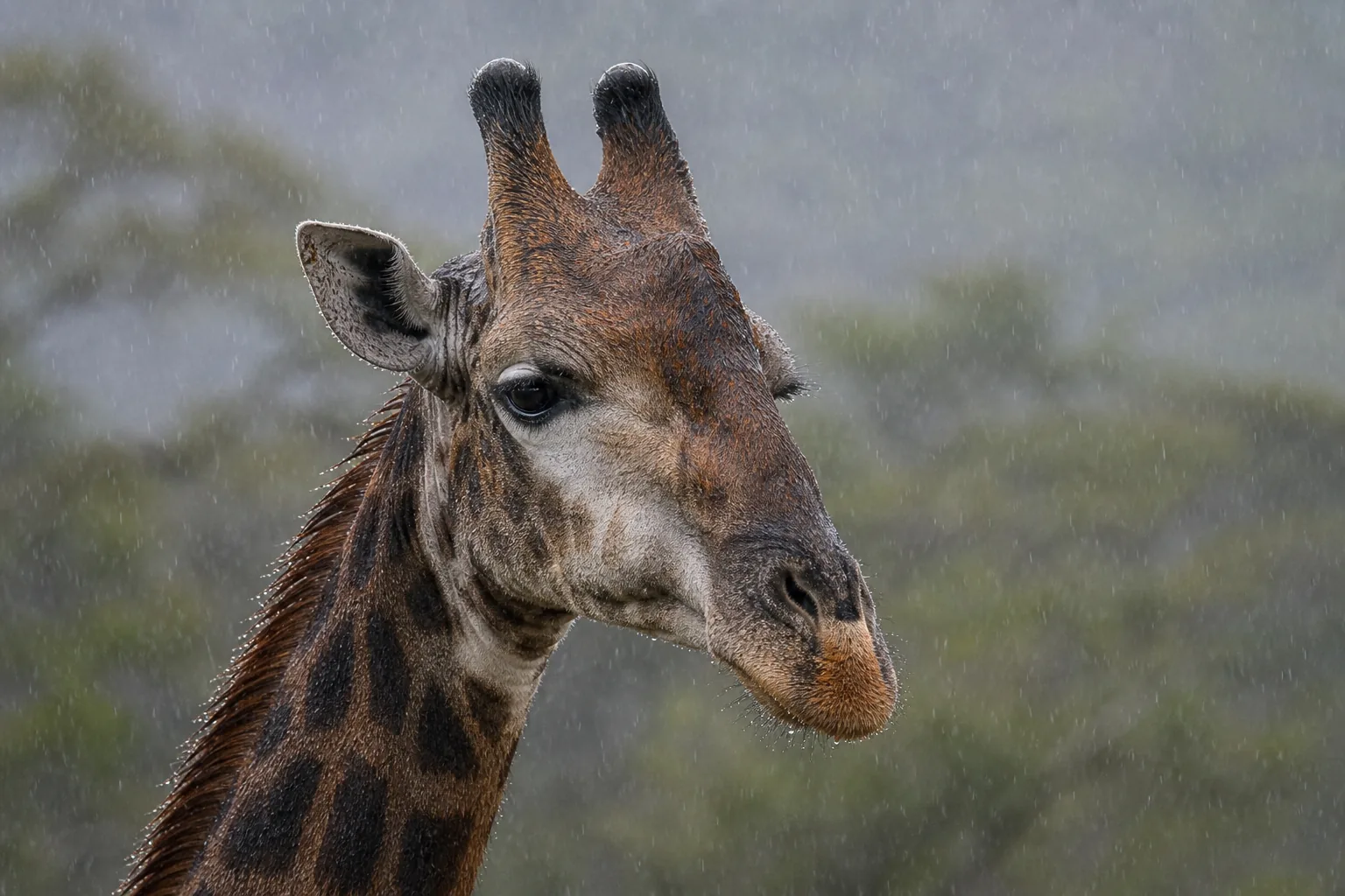
Much of the distinction involved the ossicones and the length of the muzzle. The median ossicone, a bony prominence along the middle of the forehead, was particularly informative.
In the characteristic male examples described by the authors, northern giraffes had a high, relatively pointed median prominence. Reticulated giraffes had a broader, more rounded or stepped elevation. It was smaller in Masai giraffes and faint or effectively absent in southern giraffes. Differences in this region were milder in females.
These were patterns across a variable collection. The authors explicitly noted considerable departures from the typical form within each species. A skull could belong to a statistically distinct group while resembling individuals in another group on some measurements.
That is why the study offers more than a visual identification rule. Its argument rests on combinations of three-dimensional features and comparisons across many specimens. Our ossicone guide explains the structures themselves; the new analysis examined how their form contributes to differences among giraffes.
An anatomical line of evidence alongside DNA
Genetic analyses and skull measurements describe different aspects of giraffe variation. Finding differences in both strengthens the case that the groups represent more than names attached to slightly different coat patterns.
The study nevertheless began with taxonomic assignments informed by where specimens came from. Its achievement was to test whether anatomical differences corresponded to those groupings, rather than discovering an entirely new classification without prior information.
Nor did every proposed subspecies pair separate. The two southern giraffe subspecies, Angolan and South African, were among those without a statistically significant skull-shape distinction in these comparisons. The level at which anatomy can distinguish groups need not be identical to the level at which DNA can reveal their histories.
The broader taxonomy and genetics explanation explores why classification draws on several kinds of evidence. Kargopoulos and colleagues added a large, consistently measured anatomical dataset to that discussion.
Their work also leaves a concrete developmental question: how do different growth histories produce the adult head forms? Comparisons with better age information could clarify when the differences emerge. The researchers identify the lower jaw, internal skull anatomy and the rest of the skeleton as further places to investigate. A giraffe’s head is already a recognisable silhouette; these measurements show how much biological variation lies within it.
Sources
Nikolaos Kargopoulos, Jesús Marugán-Lobón, Anusuya Chinsamy, Bernard R. Agwanda, Michael Butler Brown, Stephanie Fennessy, Sara Ferguson, Rigardt Hoffman, Fredrick Lala, Arthur Muneza, Ogeto Mwebi, Moses Otiende, Alice Petzold, Sven Winter, Abdoul Razack Moussa Zabeirou and Julian Fennessy (2024). Heads up–Four Giraffa species have distinct cranial morphology. PLOS ONE 19(12), e0315043. Read the full paper.