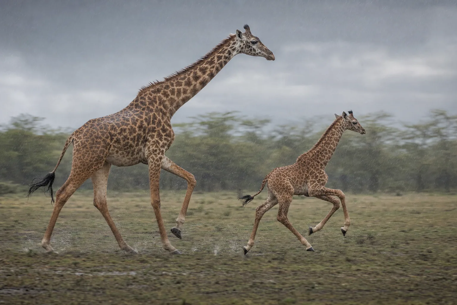
A placenta is often described as an exchange organ, carrying nutrients towards a fetus and wastes away. It also produces signals, and the cells producing them need not all perform the same job. In giraffes, a microscopic comparison revealed a striking difference between where specialised cells occurred and where particular proteins could be detected inside them.
F. B. P. Wooding and colleagues reported this pattern in a 2022 study in Biology of Reproduction. Cells of the same general type were distributed through the giraffe tissue, yet their protein reactions separated into distinct zones. The researchers found no equivalent division in the other ruminants they examined.
The calf’s tissue reaches into the mother’s
Ruminant placentas contain repeated attachment units called placentomes. Within each unit, finger-like extensions of fetal tissue fit closely with maternal tissue. These projections, or villi, provide a large interface within a compact space.
Among their specialised cells are binucleate cells, named for their two nuclei. They contain granules: small packages holding proteins that can be released. In ruminants, mature binucleate cells migrate and fuse with cells or tissue on the maternal side, allowing their contents to enter the maternal compartment.
That makes the contents of the granules biologically interesting. Their distribution is part of the machinery through which fetal and maternal tissues communicate. A cell near the base of a projection is in a different physical position from one near its tip, even if both look like the same cell type under an ordinary microscope.
Earlier giraffe observations had suggested that some protein signals might be restricted to particular locations. Wooding’s team tested the pattern more broadly by comparing preserved placental material from giraffes with that from 13 other ruminant species, including okapi and pronghorn.
Seeing a protein requires a selective label
The team used antibodies that bind to particular proteins or related molecular structures. Attached labels made those binding sites visible under the microscope. Different antibodies allowed the researchers to compare signals associated with placental lactogens, prolactin and pregnancy-associated glycoproteins, commonly shortened to PAGs.
An antibody stain is a detection method, not a direct measurement of a protein’s physiological action. Antibodies developed against one species’ proteins can react differently in another species. The researchers therefore used multiple antibodies and comparison tissues, and examined the consistency of the resulting patterns.
They also used lectins, molecules that bind to particular sugar structures. One strongly reacting lectin identified the giraffe binucleate-cell granules along the full length of the fetal projections. This supplied a crucial comparison: it showed that the cells were present even where a particular protein antibody gave little or no signal.
Without that comparison, a blank region could be mistaken for a region without the relevant cells. Instead, the study distinguished where the cells were from what their granules contained that the chosen labels could detect.
A division along the placental projection
In the giraffe samples, antibodies to placental lactogens and prolactin reacted mainly near the base of the fetal projections. Reactions to several PAG antibodies were concentrated towards the opposite, maternal-facing end. The clearest patterns occupied approximately the first and last quarters of the projection.
Cell counts reinforced what the images showed. Counts based on the broad lectin label were fairly even across the four quarters, whereas counts based on the protein-specific labels varied sharply with position. One lactogen antibody had a wider distribution than the others, so the result was a strong spatial pattern rather than an absolute rule that every detected protein occupied only one small zone.
The same broad organisation appeared in samples from two giraffes. That is a small animal sample, but it matters that the pattern was repeated and that different methods distinguished the cells from their contents. The authors also found comparable results across differently preserved tissue, reducing the likelihood that fixation alone produced the finding.
Across the other 13 species, the corresponding cells generally showed a much more even distribution of detectable granule proteins. Even the okapi did not share the giraffe’s zonation. The giraffe pattern therefore could not simply be assumed from its closest living relative.
A clear pattern with an unresolved purpose
The result adds a cellular dimension to giraffe pregnancy. Knowing that the placenta produces a signal is one level of explanation; knowing which cells contain it and where they lie is another. The paper shows that the giraffe placenta has an organisation that a whole-organ measurement could conceal.
The researchers considered whether concentrating PAGs near maternal tissue might influence the immune balance that allows fetal tissue to remain there. They also discussed the metabolic roles associated with lactogens and prolactin. These were possible explanations, not tested functions of the observed zones. The authors did not establish why giraffes use this arrangement or whether it changes delivery of signals to the mother or fetus.
Further work could identify the giraffe proteins more directly and examine whether the pattern changes as pregnancy progresses. Functional measurements would then be needed to connect their location with what they do. Larger samples would help establish how consistently the pattern occurs among individuals.
The useful discovery is already specific: cells that look like one population can be organised into different biochemical roles across a tissue. In the giraffe placenta, a map of the cells alone would have missed that distinction.
Sources
F. B. P. Wooding, A. J. Forhead, S. Wilsher, W. R. Allen, R. M. Roberts, J. A. Green, J. F. Beckers, N. Melo Sousa and G. Charpigny (2022). Asymmetric expression of proteins in the granules of the placentomal Binucleate cells in Giraffa camelopardalis. Biology of Reproduction 106(4), 814–821. DOI; open full paper.