A diet can contain a recommended amount of a nutrient while an animal still fails to obtain enough usable protection from it. The reasons may lie between the feed bowl, digestion, pregnancy and the first milk after birth. A newborn giraffe with severe muscle weakness made that distinction difficult to ignore.
Jan H. Bos, Fokko C. Klip and Marja J. L. Kik described the case in a 2017 veterinary paper. Their findings supported white muscle disease associated with low vitamin E, even though the mother’s estimated dietary supply was higher than the published giraffe feeding guidance they cited. The unresolved question was how a seemingly sufficient ration and a deficient calf could coexist.
Muscle cells need protection as well as fuel
Vitamin E helps protect cell membranes from oxidative damage. Selenium contributes to a different part of the antioxidant system through enzymes that limit damaging chemical reactions. Deficiency of either can be associated with nutritional myodegeneration: degeneration of muscle cells, traditionally called white muscle disease because affected muscle can appear pale.
The resulting weakness has a direct mechanical explanation. Damaged skeletal muscles cannot generate normal movement, and involvement of the heart or breathing muscles can make disease especially serious. A newborn struggling to stand may therefore have a problem in muscle tissue itself, rather than simply being slow to learn how to use its legs.
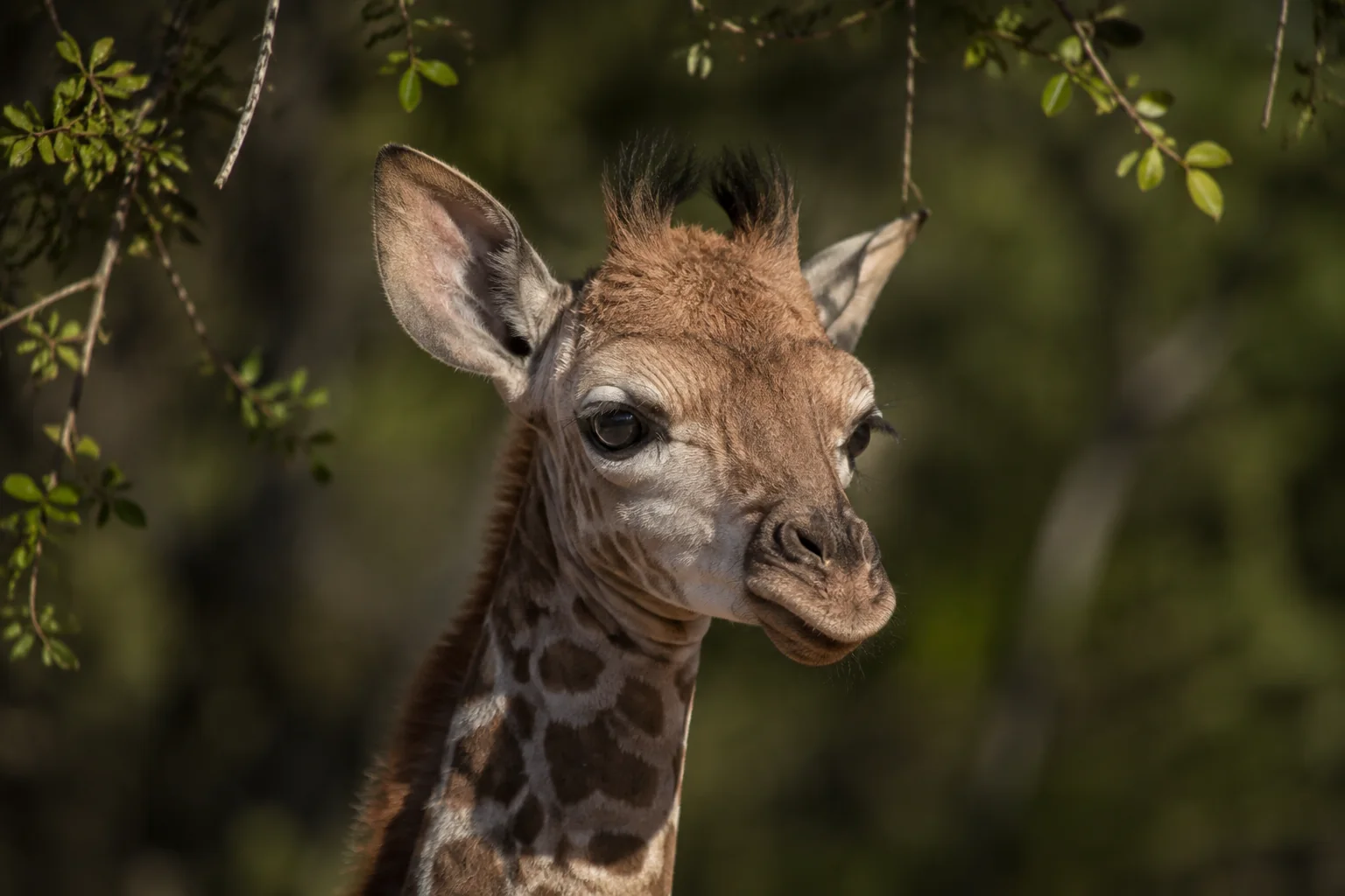
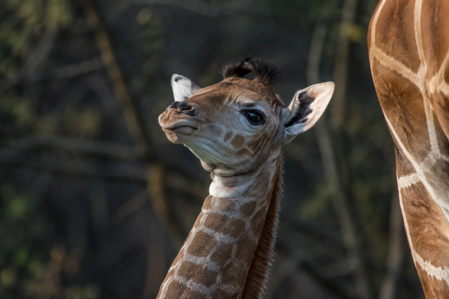
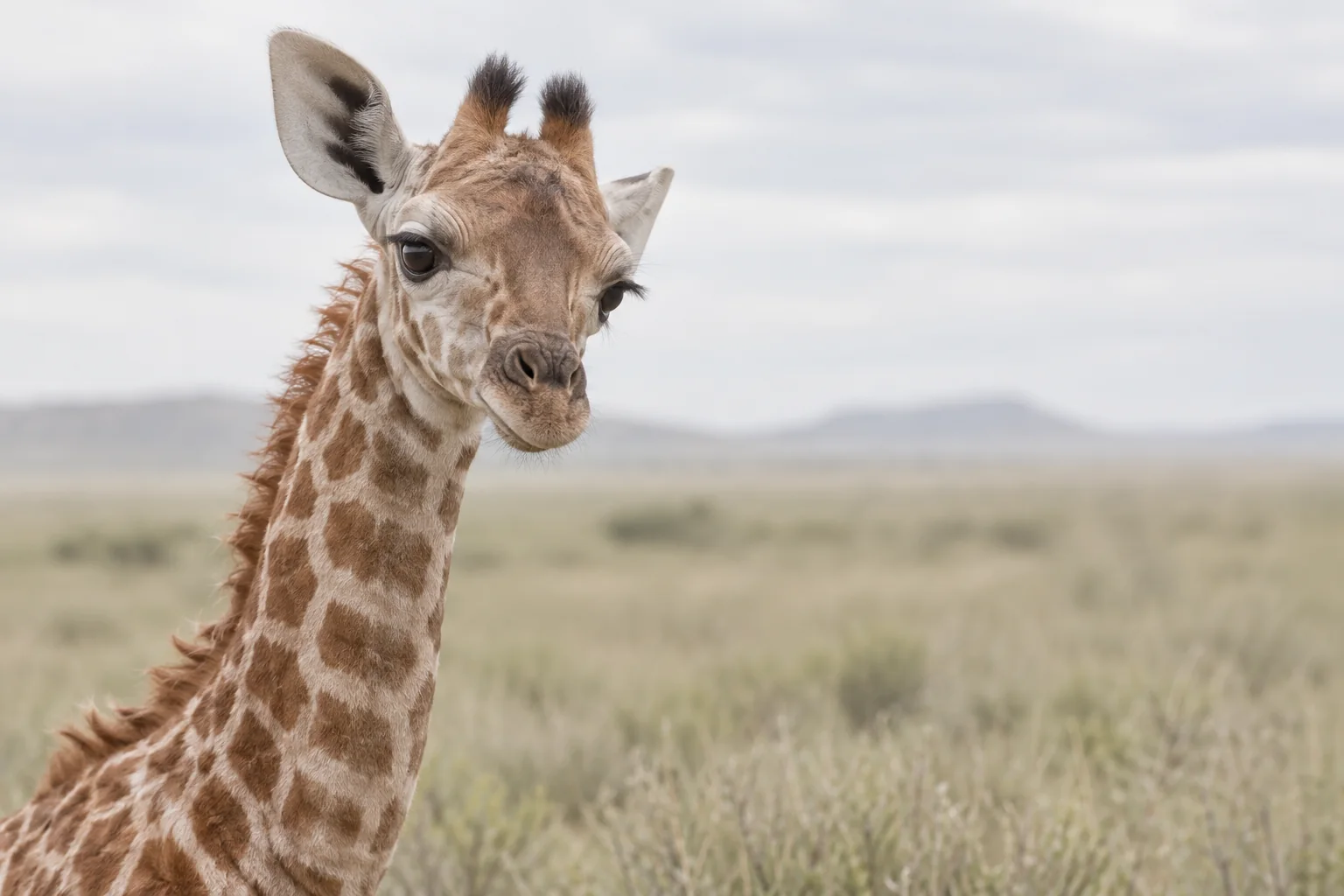
The giraffe calf was born at Ouwehand Zoo in the Netherlands. It stood within about an hour, but weakness in the hind limbs became apparent. Over the following hours, it struggled to rise and remain standing, trembled and repeatedly lay down. Its mother rejected it, and keepers began feeding stored bovine colostrum.
Despite care and supplementation, the calf deteriorated and was euthanised in its first few days. The investigators then combined blood results with a detailed examination of its tissues.
The diagnosis depended on several kinds of evidence
The calf was not simply emaciated: the necropsy described moderate to good body condition. However, its skeletal muscles and heart appeared pale, and fluid had accumulated in the lungs. Under the microscope, the sampled skeletal muscles showed degeneration of muscle cells, along with fluid accumulation and increased blood content.
Trauma around birth was among the alternative explanations the investigators considered. The blood chemistry supplied an additional clue. The first vitamin E concentration was below the laboratory’s detection limit of 0.7 mg/L. The second was 0.7 mg/L. The selenium result did not appear low against the comparisons available from other ruminants.
Taken together, the clinical weakness, widespread muscle changes and low vitamin E supported nutritional myodegeneration. The interpretation was stronger than any one observation alone: pale muscle is not a vitamin assay, and a low vitamin measurement does not show by itself which tissues have been damaged.
The comparisons were imperfect. Published blood values came from giraffes of different ages and other ruminant species, and the paper did not establish a new reference interval for healthy newborn giraffes. Its diagnosis rested on the combined case evidence, not on a population study defining a precise universal threshold.
The ration calculation looked reassuring
The researchers reviewed the mother’s diet, which consisted of lucerne hay and browser pellets. From the estimated quantities eaten and the vitamin E content of the pellets, they calculated at least about 396 international units per kilogram of dietary dry matter. The vitamin E content of the hay was unknown, so it was not included as an additional contribution.
That estimate exceeded the giraffe dietary recommendations cited in the paper, including guidance of 100–150 units per kilogram of dry matter. The authors also considered a higher recommendation for pregnant small ruminants: 5.6 international units per kilogram of body weight. Applied to this mother, that corresponded to about 396 units per kilogram of dietary dry matter, approximately the calculated ration. The estimate therefore exceeded the cited giraffe guidance, but not every relevant comparison. These historical comparisons explain the investigators’ reasoning; they do not prescribe a ration for giraffes today.
A feed calculation measures provision, however, not absorption. It also does not measure how much vitamin reaches fetal tissues or becomes available to a newborn after birth. The authors discussed uncertainty about vitamin E availability in giraffes and the limitations of applying requirements derived from domestic ruminants. The report did not establish a vitamin E requirement for newborn giraffes.
The missing maternal nursing was another possible factor. Colostrum can be important for a newborn’s vitamin supply as well as its immune protection. Yet the calf was already weak very early, and the report could not quantify the contributions of prenatal status, early milk intake or later supplementation. No single explanation was demonstrated.
What the case changed
The study did not show that a particular ration caused the disease or that increasing every giraffe’s vitamin E intake would prevent it. A single affected calf cannot establish a dietary requirement. The authors themselves concluded that the precise cause remained undetermined.
What it did show was why giraffe health investigations need to distinguish a nutrient listed in a ration from the animal’s measured status. The same distinction applies between a mother and her calf: knowing the estimated intake of one does not fully describe the biological supply reaching the other.
A useful next comparison would follow maternal vitamin status, verified feed content and calf measurements around birth in animals with different outcomes. That could help locate where provision and biological availability diverge. Bos and colleagues’ case provides the reason to ask: the feed calculation looked reassuring, while the calf’s blood and muscles told a different story.
Sources
Jan H. Bos, Fokko C. Klip and Marja J. L. Kik (2017). Congenital nutritional myodegeneration (white muscle disease) in a giraffe (Giraffa camelopardalis) calf. Journal of Zoo and Wildlife Medicine 48(4), 1193–1196. DOI; university repository PDF.