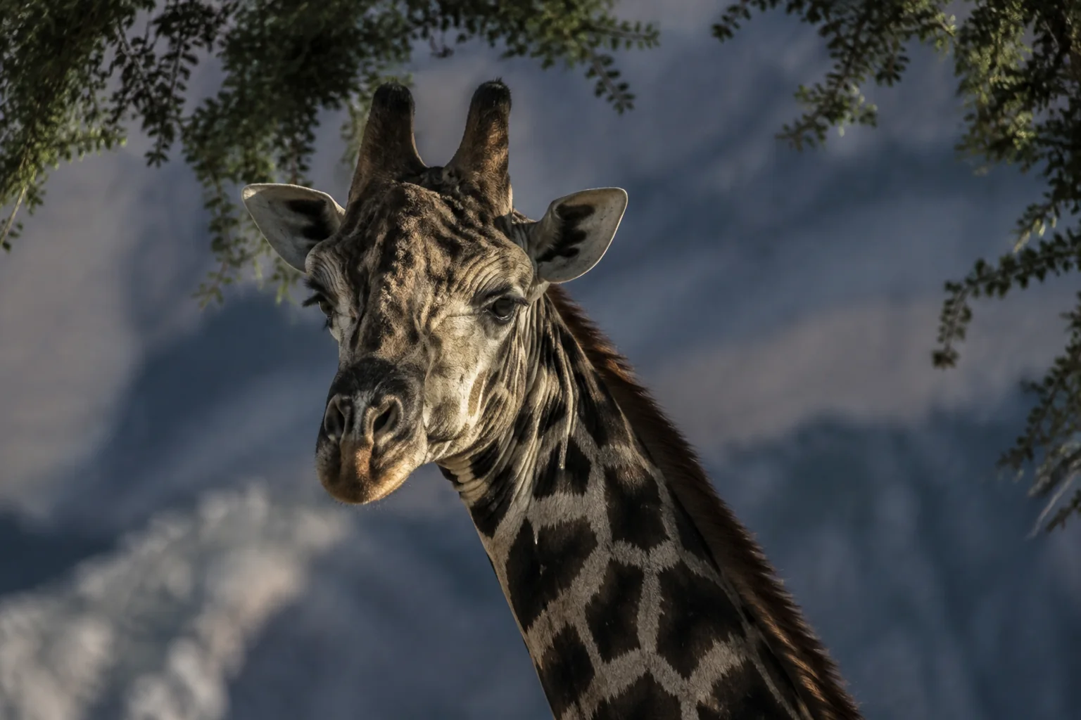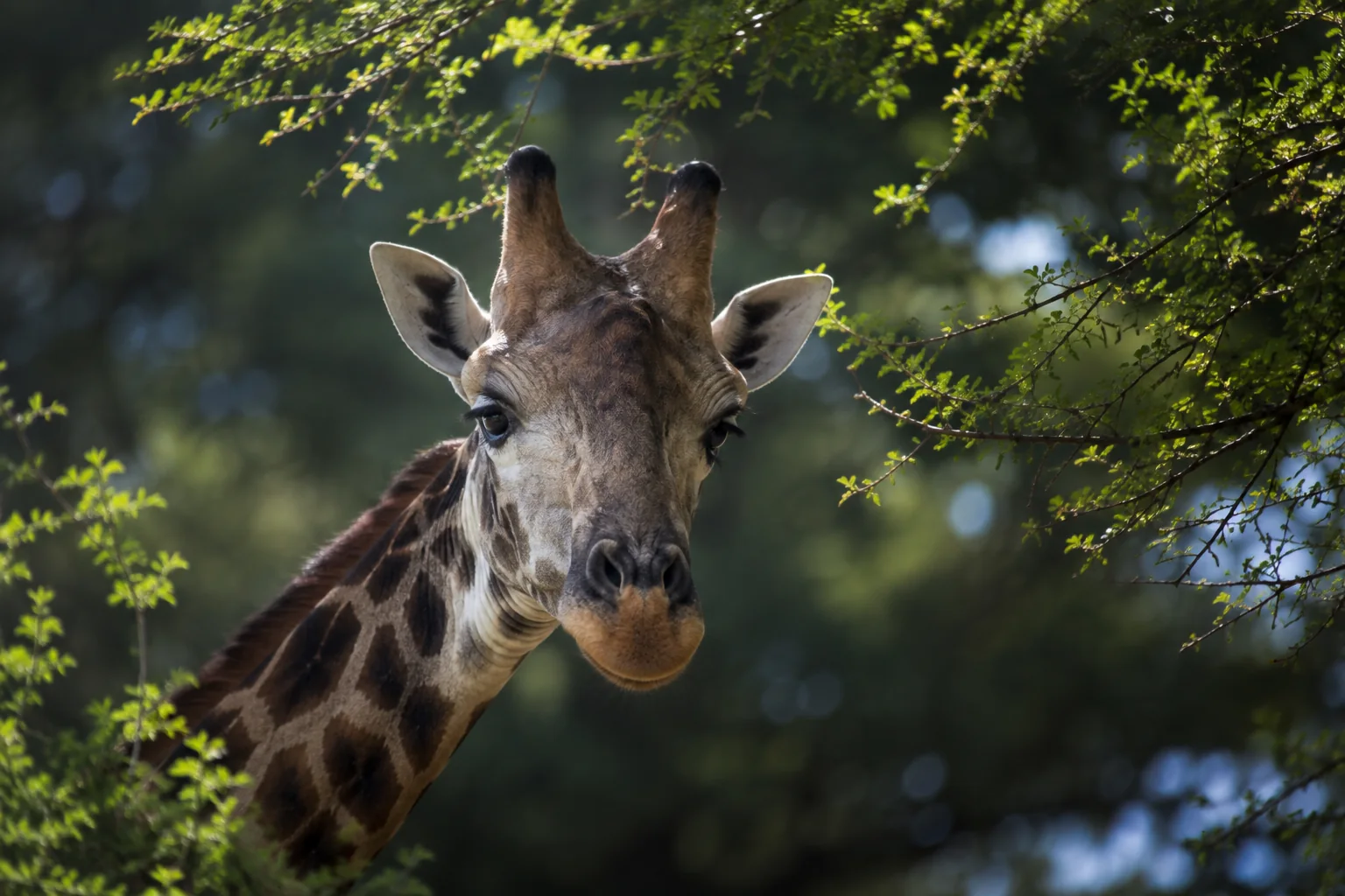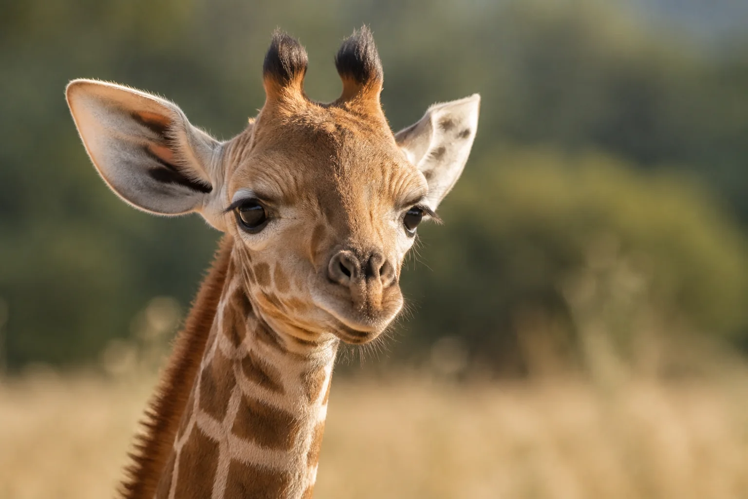
The animal in which a virus is found does not necessarily define the virus’s closest relatives. A pathogen detected in a giraffe may resemble strains better known from another host. Establishing that relationship is useful, but it answers a different question from identifying the animal that passed on the infection.
Emily Mulherin and colleagues explored this distinction in a 2008 study of a sick giraffe calf. They identified rotavirus and showed that two of its genes closely resembled those of cattle-associated viruses. The work provided a genetic description where previously the clinical signs alone could not reveal which kind of virus was involved.
From diarrhoea to visible virus particles
The orphaned Rothschild’s giraffe calf came from Dublin Zoo and was admitted to a veterinary hospital at 14 days old. It had persistent watery diarrhoea, dehydration and other serious abnormalities. The clinical history also included failure of passive immune transfer: inadequate early acquisition of protective antibodies.
The calf died four days after hospital treatment began. A postmortem examination found severe inflammation of the abomasum, the glandular stomach compartment, and the intestine. Bacterial cultures did not identify significant pathogens in the sampled organs; the researchers noted that intensive antimicrobial treatment could have affected those results.
A faecal specimen supplied a different line of evidence. Under an electron microscope, the team observed particles with the appearance and size of rotaviruses. Examining viral genetic material then confirmed the characteristic pattern of a group A rotavirus.
These observations connected the clinical disease with a recognisable pathogen. They did not by themselves establish that rotavirus was the only contributor to the calf’s death, particularly given its serious underlying vulnerabilities and the limitations of the other tests.
Two surface proteins provide a genetic address
Rotavirus carries its genetic information in 11 separate RNA segments. Two surface proteins, called VP7 and VP4, are especially useful for distinguishing strains. The genes encoding them form the basis of a two-part classification: a G type and a P type.
The team amplified these genes from the giraffe sample, obtaining a complete sequence for VP7 and a partial sequence for VP4. That difference is important. The study described targeted parts of the virus, not a complete genome assembled from all 11 segments.
The resulting classification was G10P[11]. Rather than leaving that label as an unexplained code, the researchers compared the sequences with reference strains from several animal hosts. Both comparisons placed the giraffe virus close to cattle-associated strains. The paper reported about 98% similarity at the amino-acid level in the relevant sequence comparisons.
The genetic trees provide the reasoning behind the result. They group sequences according to their differences, asking which previously characterised viruses are most similar in the regions examined. The giraffe sample’s placement was evidence of relatedness. It was not a diagram of animals infecting one another.
Relatedness does not identify the source
The calf reportedly had no direct contact with other ruminants before arriving at the veterinary hospital. That made it especially important not to translate a cattle-like sequence into a claim that a cow had infected it.
The researchers considered possible indirect contacts through staff movements, feeding equipment and cleaning equipment. These were routes worth investigating, not demonstrated links. There were no matched samples establishing a chain from a particular animal, enclosure or item of equipment to the giraffe.
A further complication is that a virus type can circulate in more than one host. A strain described as cattle-associated may also be detected elsewhere. Without sampling the relevant animals and environment at the relevant time, a genetic resemblance cannot locate the source.
That is why the study’s strongest conclusion was about the virus’s identity and relationships. It broadened the known host context for this genetic type while leaving the actual infection route unresolved.
Why the separate RNA segments matter
When different rotaviruses infect the same cell, their genome segments can sometimes be mixed into new combinations. This process, called reassortment, is one reason scientists monitor more than a single viral feature across hosts.
The calf study did not demonstrate a reassortment event. Its two-gene analysis could establish the G and P types and their closest comparisons, but the remaining segments would need to be examined to describe the whole genetic combination. More comprehensive sequencing could test whether all parts of a virus share the same apparent history.
For giraffe health research, the work shows why identifying a pathogen beyond its broad family can be valuable. “Rotavirus was present” and “these genes resemble cattle-associated strains” are different levels of knowledge. The second can guide which related hosts and virus populations to investigate, provided it is not mistaken for proof of transmission.
Mulherin and colleagues supplied that more specific description from a difficult clinical case. The remaining question was no longer simply what the particles were, but how viruses with that genetic profile were moving through the wider animal setting.
Sources
Emily Mulherin, Jill Bryan, Marijke Beltman, Luke O’Grady, Eugene Pidgeon, Lucie Garon, Andrew Lloyd, John Bainbridge, Helen O’Shea, Paul Whyte and Séamus Fanning (2008). Molecular characterisation of a bovine-like rotavirus detected from a giraffe. BMC Veterinary Research 4, 46. DOI; open full paper.