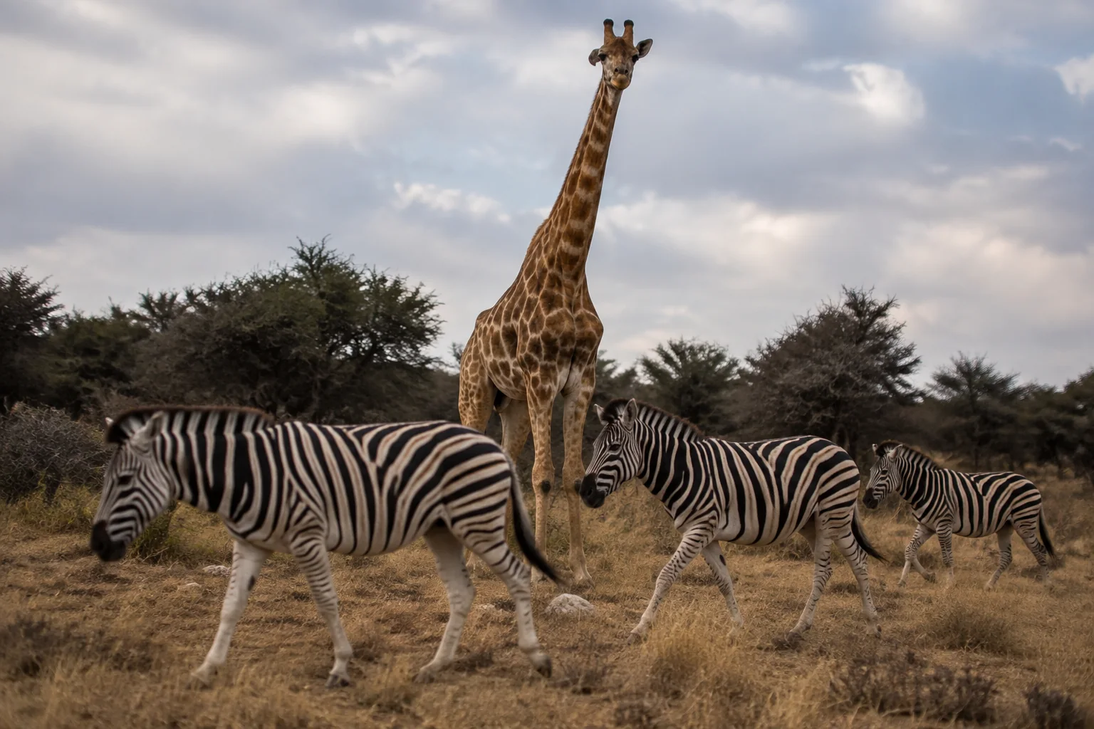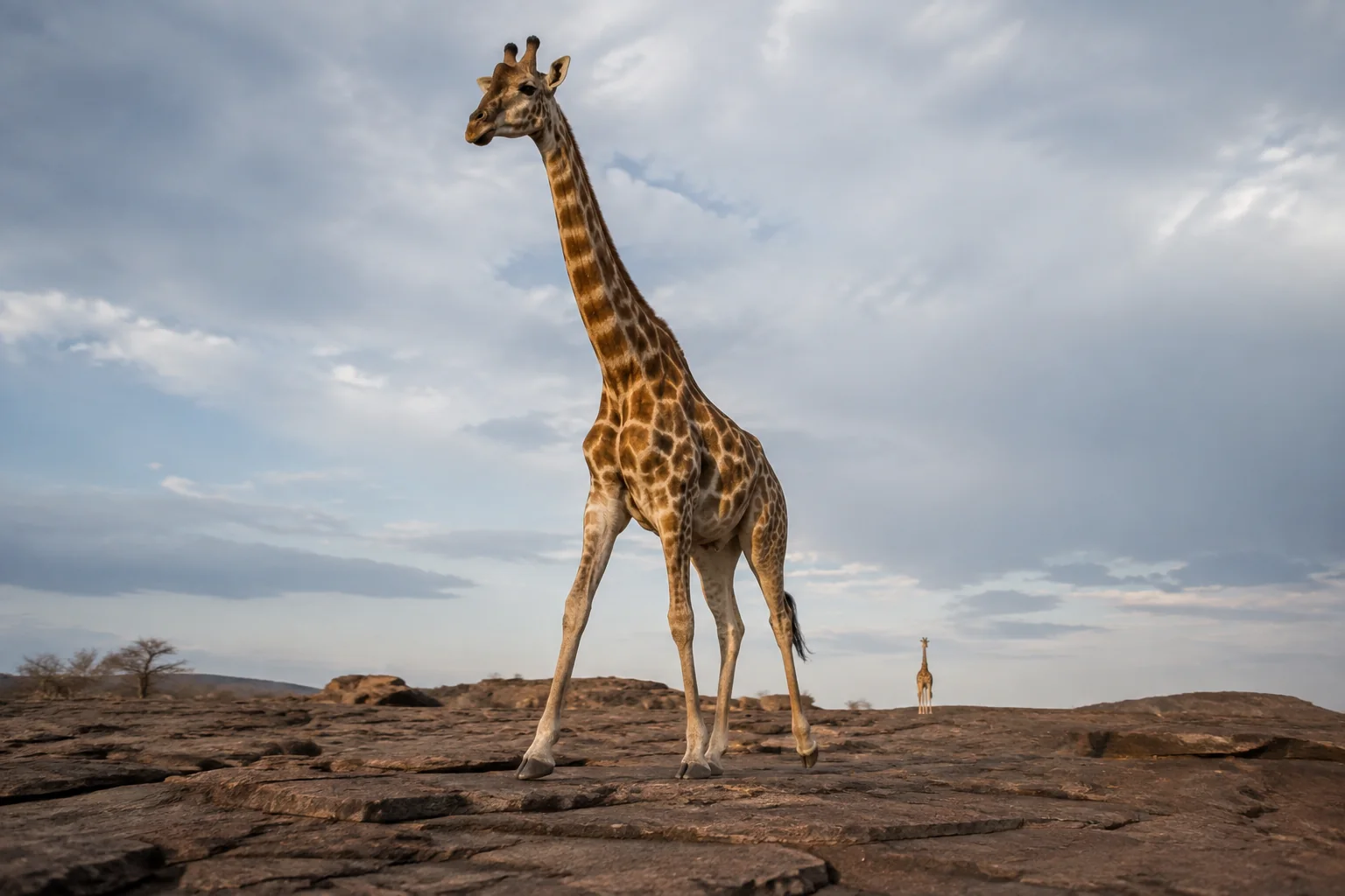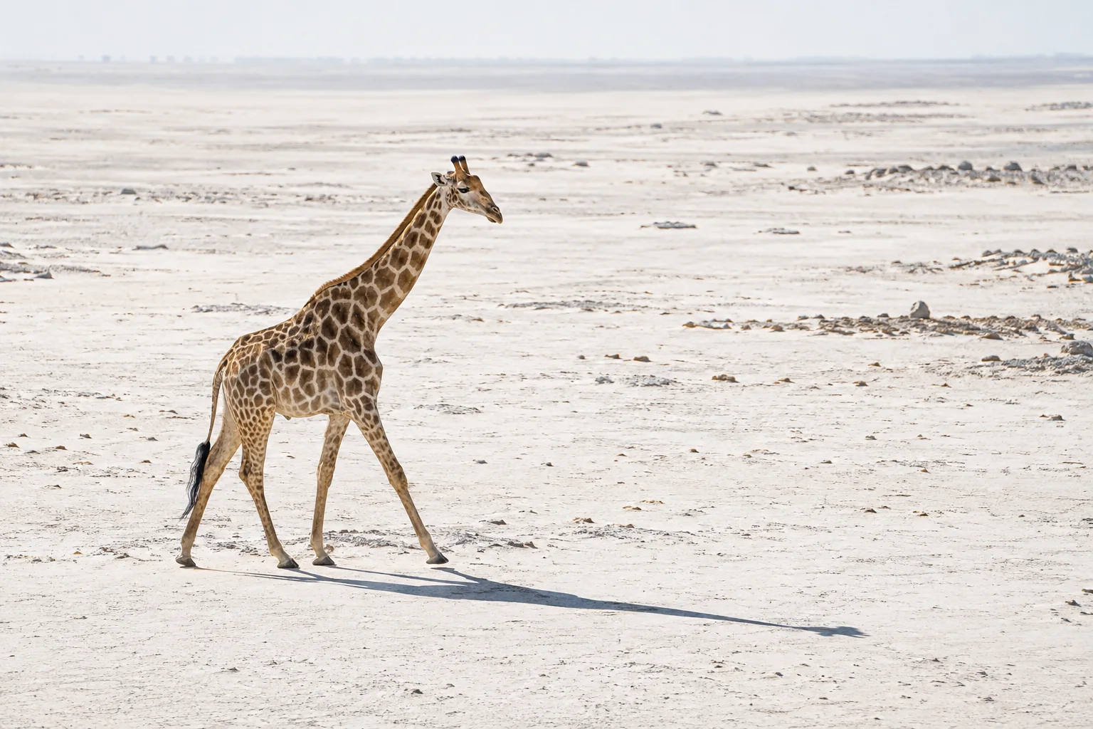
A large abdomen can mean several different things. It may reflect a large skeleton, the contents of a digestive system, pregnancy or stored tissue. For a giraffe, even obtaining a reliable record of its external shape is difficult: an observer sees a very large animal from a limited set of angles, and a suitable weighing platform may be unavailable.
A 2024 study by Nobuhide Kido and colleagues asked whether handheld laser measurements could turn that outline into a set of comparable numbers. The team scanned seven giraffes, five okapis and three black rhinoceroses in three Japanese zoos. They could describe how each trunk widened and narrowed, and estimate the volume enclosed by part of it.
The most informative result was that different ways of describing shape did not always tell the same story. An animal with a relatively bulging abdomen was not necessarily the one with the greatest trunk volume for its length. That distinction is central to making the technique useful: the surface can be measured before researchers know exactly which measurement best indicates biological condition. Read the focal study.
Turning a surface into a series of sections
Two operators recorded the animals from opposite sides, using handheld instruments connected to computers. Each image took about a minute to acquire. Food helped the animals remain in position without sedation or special training for the procedure.
The resulting data formed a cloud of points representing the visible surface. Software converted those points into a model from which the researchers could take virtual sections across the trunk. They measured each section’s width, height and enclosed area at five-centimetre intervals, starting near the armpits and continuing toward the groin.
A section’s area contains information that width alone cannot provide. A trunk can become wider but shallower, leaving its total cross-sectional area little changed. The measurements therefore separated two dimensions that a single side view can easily combine into one impression of a “large” abdomen.
Not every part of the surface was recorded. Where scans had gaps, especially underneath the abdomen, the researchers estimated the missing outline using data from other individuals of the same species. The completed model was consequently a mixture of measured points and reconstructed surface. That distinction matters whenever a small apparent difference might depend on an unseen region.
Using the animal’s own chest as a reference
The first comparison expressed each section relative to the section near the armpits. If that starting area was assigned a value of 100%, a later section at 120% represented an area one-fifth larger. This described the way the animal’s trunk changed along its length, rather than simply identifying which animal was largest.
Three giraffes had a noticeable mid-trunk expansion in area relative to their own starting section. Other giraffes did not show that pattern. All seven nevertheless widened around the middle to some degree. Their body depth tended to decrease toward the rear, helping explain why width and area were not interchangeable.
The scans also showed differences within the okapi and rhinoceros groups. That established that the procedure could recover a range of trunk outlines across animals with very different overall forms. It did not establish a common healthy shape that could be applied to all three species.
A ratio brings its own interpretive problem. An animal with a large starting chest can have a substantial abdomen without a dramatic increase relative to that chest. A more cylindrical trunk may produce a comparatively flat profile even when its absolute volume is large.
A second comparison exposed the limits of the first
The researchers also estimated the volume between the armpit and groin regions. They added the contributions of successive sections, assuming that area changed linearly between them. This was an approximate trunk volume, excluding much of the neck, limbs and head. It was not a measured whole-body weight.
They then compared that volume with body length. Broadly, longer animals within each species had larger estimated volumes, but individual positions mattered. Giraffe No. 2, for example, had a relatively large volume for its length without the pronounced relative abdominal expansion identified in some other giraffes.
The two results need not contradict each other. One asks how the abdomen compares with the animal’s own front section. The other asks how much space the measured trunk encloses in relation to overall length. Those are different anatomical questions.
The distinction is particularly important for an animal that is pregnant. One giraffe in the study was pregnant, and its measurements could not simply be interpreted in the same way as those of an otherwise similar non-pregnant individual. Recent feeding can also alter the volume of digestive contents in giraffes and okapis. Neither effect is a direct measure of stored fat.
What the earlier giraffe experiment had already revealed
The team’s 2018 study used the same general technique on three giraffes. Ten zoo staff also scored their body condition visually. The animal receiving the highest average visual score was not the one with the largest measured cross-sectional area and width relative to body length.
That disagreement showed that the methods captured different information; it did not prove that the scanner had identified the fattest animal. No direct measurement of fat resolved the comparison. A basic accuracy check also left work to do: one giraffe’s chest circumference measured 220 centimetres with a tape, compared with approximately 253 centimetres in the digital model. The authors considered differences in measurement location and measurement error possible explanations. Read the earlier experiment.
The later study extended scanning to more individuals and species and compared relative shape with approximate volume. It still did not provide an independent body-composition measurement against which to validate a nutritional diagnosis. The earlier discrepancy therefore remains relevant when judging what the newer measurements establish.
A useful record, with a validation step still ahead
The immediate achievement is a quantitative description of an animal’s exterior. Such a record could support comparisons that are hard to make from memory or a single photograph. But this study scanned the animals once; it did not demonstrate how reliably repeated scans detect gradual changes in the same individual.
A stronger biological interpretation would require repeated measurements at consistent positions, checks on reconstructed areas and comparisons with independent evidence of body mass or tissue stores. It would also need to account for maturity, pregnancy and feeding state. These are practical questions about which differences the method is detecting.
The study makes the next step clearer because its two shape measures sometimes disagree. Rather than selecting whichever number looks most decisive, researchers can ask why the numbers diverge and which one tracks the biological change of interest. A detailed outline offers a starting measurement. Establishing what that outline means requires a separate comparison with the animal beneath it.
Sources
Nobuhide Kido, Sohei Tanaka, Yuko Wada, Atsushi Oura, Emi Ochiai, Natsumi Morita, Yoshiya Kawaguchi, Masanori Itabashi and Takanori Munakata (2024). “Body Shape Analysis in Reticulated Giraffe, Okapi, and Black Rhinoceros Using Three-Dimensional Laser Measurements.” Journal of Zoological and Botanical Gardens 5: 80–89. Full paper.
Nobuhide Kido, Sohei Tanaka, Tomoko Omiya, Yuko Wada, Mina Shigenari, Takanori Munakata and Masaki Ogawa (2018). “Evaluation of somatotype in the reticulated giraffe (Giraffa camelopardalis reticulata) using three-dimensional laser measurement.” Journal of Veterinary Medical Science 80: 1528–1533. Full paper.